New Nanoparticles Illuminate and Destroy Hidden Brain Cancer in Mice, Offering Hope for Glioblastoma Patients
Introduction
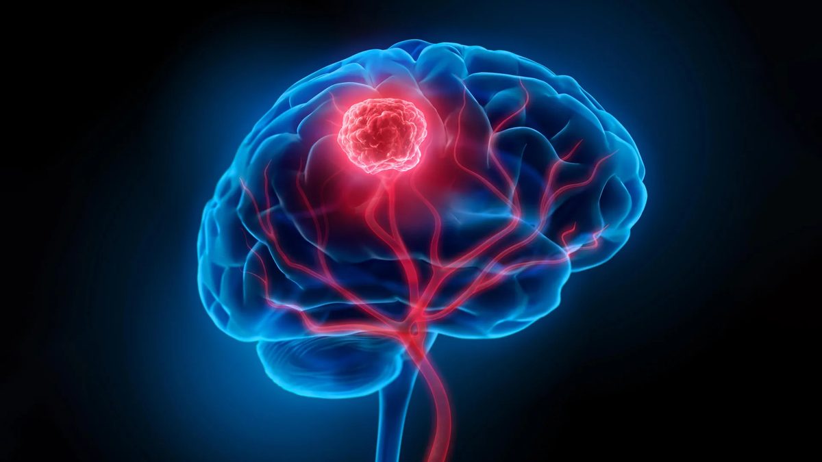
Brain cancer, particularly the aggressive form known as glioblastoma, presents one of medicine's most formidable challenges. The diffuse nature of cancer cells, which infiltrate surrounding healthy brain tissue, makes complete surgical removal a perilous endeavor. Surgeons must navigate a delicate balance, aiming to excise as much of the tumor as possible while preserving vital neurological functions. Compounding this difficulty is the blood-brain barrier, a biological shield that severely limits the efficacy of conventional drug and radiation therapies. These combined obstacles contribute to the grim reality of glioblastoma, where the five-year survival rate hovers at a mere 7 percent. However, a groundbreaking development from researchers at the University of Technology Sydney (UTS), Harvard University, and Henan University offers a glimmer of hope, introducing a novel nanoparticle platform designed to tackle these issues head-on.
Key Details
- Dual-Function Nanoparticles: Researchers have engineered a 'double-punch' nanozyme platform using smart nanoparticles capable of both imaging and treating brain cancer.
- Enhanced Surgical Visualization: The nanoparticles, when activated by near-infrared light, fluoresce, allowing surgeons to detect individual tumor cell clusters as small as 44 micrometers, significantly improving upon current imaging capabilities.
- Targeted Post-Operative Therapy: After visible tumor removal, the same nanoparticles are used to destroy microscopic cancer cells left behind. They convert the tumor's own hydrogen peroxide into oxygen, counteracting a protective low-oxygen environment, while light activation generates heat and reactive molecules to eliminate residual cancer.
- Improved Survival Rates in Mice: In mouse models of glioblastoma, this nanoparticle treatment resulted in 100% survival at 60 days, compared to 42 days for surgery alone, and significantly reduced tumor recurrence.
- Blood-Brain Barrier Penetration: A targeting molecule attached to the nanoparticles facilitates their passage across the blood-brain barrier, enabling them to accumulate specifically in glioma cells.
Background
Glioblastoma's notorious reputation stems from its rapid growth and tendency to spread stealthily within the brain. Unlike more contained tumors, its tendrils can extend far beyond the visible margins, making surgical resection a race against unseen invaders. The inherent complexity of brain anatomy further complicates matters, as aggressive surgical intervention risks causing irreversible damage to critical cognitive and motor functions. The blood-brain barrier, a highly selective membrane, acts as a formidable gatekeeper, preventing many therapeutic agents from reaching cancerous cells within the brain. This dual challenge of incomplete surgical removal and limited drug penetration has historically led to poor patient outcomes.
Impact Analysis
The newly developed nanoparticle platform, detailed in the journal Science Translational Medicine, represents a significant leap forward by addressing both the visualization and treatment gaps simultaneously. Dr. Bingyang Shi, a leading nanomedicine expert at UTS and a key author of the study, explained the innovation: “We've engineered a single material that does two jobs in sequence. It's a precise guide for the surgeon during the operation, and then a targeted clean-up treatment afterward.” The ability to visualize minuscule tumor clusters, down to 44 micrometers, is particularly revolutionary. This level of detail surpasses current clinical imaging tools, potentially enabling surgeons to achieve more complete resections without compromising healthy tissue. Furthermore, the post-operative phototherapy, which leverages the tumor's microenvironment and external light activation, offers a novel way to eliminate residual disease, a primary driver of recurrence.
“The results are very encouraging, but this is still early-stage research carried out in mouse models, not in people - and that distinction is important,” said Professor Shi. “Its imaging and therapeutic performance will also need to be confirmed at the scale of a human brain.”
Broader Context
The development of 'smart' nanomaterials for medical applications is a rapidly expanding field. These engineered particles, often thousands of times smaller than a human hair, can be designed to carry drugs, enhance imaging, or even perform therapeutic functions. This particular platform's ability to switch between imaging and therapeutic modes, activated by the same external stimulus (near-infrared light), showcases sophisticated engineering. The use of 2D materials, akin to those used in semiconductor manufacturing, highlights the interdisciplinary nature of modern scientific advancement. By converting the tumor's own hydrogen peroxide into oxygen, the nanoparticles not only create a more conducive environment for treatment but also counteract a known mechanism by which cancer cells evade therapy. This intelligent design addresses multiple facets of glioblastoma's resistance to treatment.
Future Outlook
While the results in mouse models are exceptionally promising, demonstrating 100% survival and reduced recurrence, the path to clinical application requires further rigorous investigation. The researchers are focused on validating these findings in larger animal models and eventually in human clinical trials. Key challenges will include scaling up production of the nanoparticles, ensuring their safety and efficacy in the complex human system, and confirming their performance in the human brain. If successful, this technology could fundamentally change the surgical management of glioblastoma, offering patients a better chance at long-term survival and improved quality of life by minimizing tumor recurrence. The potential for such 'smart' theranostic (combining therapy and diagnostics) agents extends beyond brain cancer, offering a blueprint for tackling other challenging diseases.
Conclusion
The development of these dual-function smart nanoparticles represents a significant scientific achievement in the fight against glioblastoma. By providing surgeons with unprecedented visualization capabilities and offering a targeted post-operative therapy, this technology has shown remarkable success in preclinical mouse models. The 100% survival rate observed is a powerful indicator of its potential. While human trials are the crucial next step, this innovation underscores the transformative power of nanotechnology in medicine and offers renewed hope for patients facing one of the most aggressive forms of cancer.
Source: sciencedaily.com